-
Depositing Lab
-
Depositing OrganizationNational Institute of Child Health and Human Development (NICHD)
Located in the United States of America -
Publication
-
Citations
-
Sequence Information
Ordering
| Item | Catalog # | Description | Quantity | Price (USD) | |
|---|---|---|---|---|---|
| Plasmid | 11912 | Standard format: Plasmid sent in bacteria as agar stab | 1 | $94 | |
| DNA | 11912-D50 | 50 μg of DNA in Tris buffer | $495 | ||
| DNA | 11912-D100 | 100 μg of DNA in Tris buffer | $585 | ||
Backbone
-
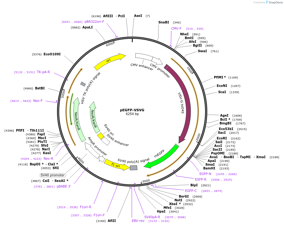
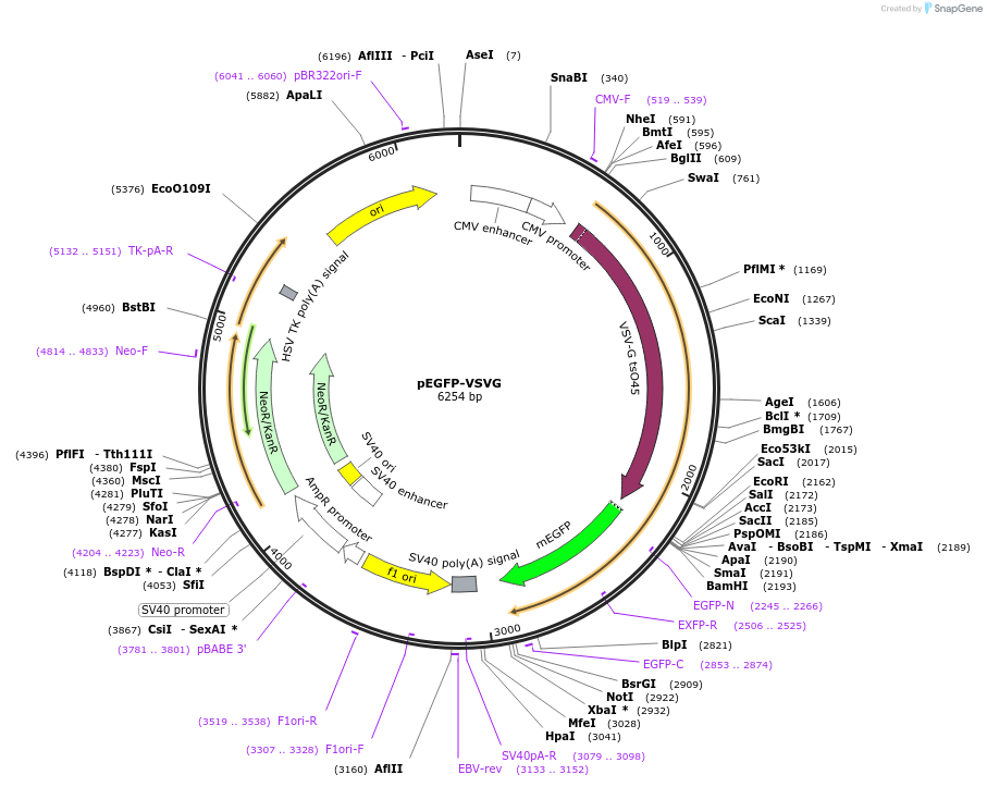
Vector backbonepEGFP-N1
-
Backbone manufacturerClontech
- Backbone size w/o insert (bp) 4733
-
Vector typeMammalian Expression
-
Selectable markersNeomycin (select with G418)
Growth in Bacteria
-
Bacterial Resistance(s)Kanamycin, 50 μg/mL
-
Growth Temperature37°C
-
Growth Strain(s)DH5alpha
-
Copy numberUnknown
Gene/Insert
-
Gene/Insert nameVSVG
-
Alt nameVesicular stomatitus virus G protein
-
Insert Size (bp)1500
-
MutationF64L, S65T, A206K relative the wild type GFP sequence. The EGFP (Clontech Laboratories, Inc., Palo, Alto, CA) contains a valine immediately after the start codon that is not found in the wild type sequence. However, to avoid confusion with previously published work on GFP mutants, the wild type residue numbers are used.
Cloning Information
- Cloning method Restriction Enzyme
- 5′ cloning site BglII (not destroyed)
- 3′ cloning site EcoRI (not destroyed)
- 5′ sequencing primer CMV-F
- (Common Sequencing Primers)
Terms and Licenses
-
Academic/Nonprofit Terms
-
Industry Terms
- Not Available to Industry
Trademarks:
- Zeocin® is an InvivoGen trademark.
Depositor Comments
The EGFP (Clontech Laboratories, Inc., Palo, Alto, CA) contains a valine immediately after the start codon that is not found in the wild type sequence. However, to avoid confusion with previously published work on GFP mutants, the wild type residue numbers are used. The Kozak sequence at the beginning of the GFP encoding region of the pEGFP-N1 plasmid from Clontech has been disrupted. Reference for the A206K mutation is Zacharias, D. A., Violin, J. D., Newton, A. C., and Tsien, R. Y. (2002) Science 296, 913-6.
DNA (Catalog # 11912-D50)
Purpose
Ready-to-use, high-purity DNA prep. DNA aliquots are suitable for use in mammalian cell transfections and other molecular biology applications.
Delivery
- Amount 50 μg
- Concentration 1 μg/μL
- Pricing $495 USD
- Storage 4 ℃ (short-term) or -20 ℃ (long-term)
Terms and Licenses
-
Academic/Nonprofit Terms
-
Industry Terms
- Not Available to Industry
Quality Control
Every DNA prep is verified by next-generation sequencing, confirming plasmid identity, sequence integrity, and absence of DNA contaminants. Concentration is confirmed to be within ±2.5% of the stated concentration.
Our production process has been validated to consistently yield endotoxin levels below 1.0 EU/μg. Individual lots are not tested for endotoxin.
DNA (Catalog # 11912-D100)
Purpose
Ready-to-use, high-purity DNA prep. DNA aliquots are suitable for use in mammalian cell transfections and other molecular biology applications.
Delivery
- Amount 100 μg
- Concentration 1 μg/μL
- Pricing $585 USD
- Storage 4 ℃ (short-term) or -20 ℃ (long-term)
Terms and Licenses
-
Academic/Nonprofit Terms
-
Industry Terms
- Not Available to Industry
Quality Control
Every DNA prep is verified by next-generation sequencing, confirming plasmid identity, sequence integrity, and absence of DNA contaminants. Concentration is confirmed to be within ±2.5% of the stated concentration.
Our production process has been validated to consistently yield endotoxin levels below 1.0 EU/μg. Individual lots are not tested for endotoxin.
These plasmids were created by your colleagues. Please acknowledge the Principal Investigator, cite the article in which the plasmids were described, and include Addgene in the Materials and Methods of your future publications.
-
For your Materials & Methods section:
pEGFP-VSVG was a gift from Jennifer Lippincott-Schwartz (Addgene plasmid # 11912 ; http://n2t.net/addgene:11912 ; RRID:Addgene_11912) -
For your References section:
ER-to-Golgi transport visualized in living cells. Presley JF, Cole NB, Schroer TA, Hirschberg K, Zaal KJ, Lippincott-Schwartz J. Nature. 1997 Sep 4. 389(6646):81-5. 10.1038/38001 PubMed 9288971