

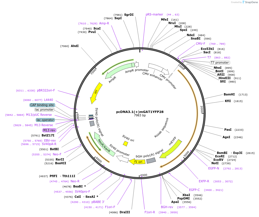
pcDNA3.1(+)mGAT1YFP28
(Plasmid
#41673)
-
Depositing Lab
-
Publication
-
Sequence Information


Ordering
| Item | Catalog # | Description | Quantity | Price (USD) | |
|---|---|---|---|---|---|
| Plasmid | 41673 | Standard format: Plasmid sent in bacteria as agar stab | 1 | $94 | |
Backbone
-
Vector backbonepcDNA3.1(+)
-
Backbone manufacturerInvitrogen
- Backbone size w/o insert (bp) 5428
- Total vector size (bp) 7983
-
Vector typeMammalian Expression
-
Selectable markersNeomycin (select with G418)
Growth in Bacteria
-
Bacterial Resistance(s)Ampicillin, 100 μg/mL
-
Growth Temperature37°C
-
Growth Strain(s)Top10
-
Copy numberHigh Copy
Gene/Insert
-
Gene/Insert nameMus musculus GABA transporter 1
-
Alt namemGAT1
-
Alt namesolute carrier family 6 member 1
-
Alt namesodium- and chloride-dependent GABA transporter 1
-
SpeciesM. musculus (mouse), Synthetic
-
Insert Size (bp)2634
-
Mutation“Monomerizing” YFP A206K mutation; 28 C-terminal-most residues of hGAT1 (QPSEDIVRPENGPEQPQAGSSTSKEAYI) appended after YFP
-
GenBank IDM92378.1
-
Entrez GeneSlc6a1 (a.k.a. A730043E01, GABATHG, GABATR, GAT-1, Gabt, Gabt1, Gat1, XT-1, Xtrp1)
- Promoter CMV
-
Tag
/ Fusion Protein
- YFP (C terminal on insert)
Cloning Information
- Cloning method Restriction Enzyme
- 5′ cloning site HindIII (not destroyed)
- 3′ cloning site XbaI (not destroyed)
- 5′ sequencing primer CMV-F; T7
- 3′ sequencing primer EGFP-N; BGH-rev
- (Common Sequencing Primers)
Terms and Licenses
-
Academic/Nonprofit Terms
-
Industry Terms
- Not Available to Industry
Trademarks:
- Zeocin® is an InvivoGen trademark.
Depositor Comments
To generate the fluorescent mutants mGAT10XFP and mGAT1XFP* through mGAT1XFP45, the wild-type mGAT1 open reading frame (ORF) was subcloned without its original stop codon into the HindIII and EcoRI sites of the pcDNA3.1(+) expression vector multiple cloning site (MCS). XFP ORFs were then subcloned downstream from and in frame with the mGAT1 ORF at the NotI and XbaI sites of the pcDNA3.1(+) MCS. This resulted in a 12–amino acid spacer between the end of the mGAT1 sequence and the beginning of the fluorophore. The depositors modified a method for the integration of PCR fragments without the use of restriction enzymes (Geiser et al., 2001) to add the final 3, 8, 20, 28, or 45 codons of the human GAT1 (hGAT1) ORF. These were amplified from a source plasmid using the proof-reading PfuTurbo Cx Hotstart polymerase with 5′ and 3′ extensions corresponding to the 20–22-nt regions that flanked the intended site of insertion, such that the PCR product integrated in-frame immediately after the fluorophore sequence when used as the primers in a subsequent QuikChange II XL mutagenesis PCR reaction. For mGAT1XFP*, the depositors simply added a GTC codon for Val after the fluorophore ORF.
The attached image displays the protein sequences of the modified regions of mGAT1 for each fluorescent construct. mGAT10CFP and mGAT10YFP repeated the fusion design of mGAT10GFP but with the fluorophore exchanged as annotated. The three C-terminal residues of the mGAT0XFP fusions are -YKI-CO2−, which comprises a broadly defined consensus PDZ class II–interacting motif (X-φ-X-φ, where φ designates a hydrophobic residue and X any residue) (Sheng and Sala, 2001; Hung and Sheng, 2002). The depositors searched the Ensembl databases using Biomart (http://www.ebi.ac.uk/biomart) (Spudich et al., 2007) and applied the GO:0005886 “plasma membrane” cellular component filter. The search identified no known membrane proteins possessing the -YKI-CO2− C-terminal sequence.
In the mGAT1XFP* constructs, the depositors defined the terminal residue P(0) more narrowly, changing the terminal isoleucine residue present in mGAT10XFP to a valine in mGAT1XFP*. The resulting C-terminal sequence, -YKV-CO2−, reconstituted a functional PDZ class II–interacting motif present in Ephrin B receptors, a class that relies on interactions with the PDZ domain–containing proteins for clustering (Torres et al., 1998; Brückner et al., 1999; Lin et al., 1999; Madsen et al., 2005).
Other constructs in the C-terminal XFP fusion series, mGAT1XFP3, mGAT1XFP8, mGAT1XFP20, mGAT1XFP28, and mGAT1XFP45, had the most C-terminal 3, 8, 20, 28, or 45 residues of the hGAT1 appended after the mGAT1XFP fusion. The differences in nucleotide sequence between the hGAT1 and mGAT1 C termini were a useful source of positive identification when the depositors analyzed the clones during construction.
PCR integration was applied to amplify and insert EYFP or ECFP directly between residues R565 and L566, I570 and Q571, or V577 and R578 of mGAT1 to generate the mGAT15xxXFP5xxCT constructs. The site of XFP insertion in GAT1 is highlighted in the nomenclatures for these constructs by residue numbers flanking the fluorophore, and the “CT” denotes that the insertion occurs within the C terminus.
Please see the associated article for more detailed information regarding construct creation and usage.
These plasmids were created by your colleagues. Please acknowledge the Principal Investigator, cite the article in which the plasmids were described, and include Addgene in the Materials and Methods of your future publications.
-
For your Materials & Methods section:
pcDNA3.1(+)mGAT1YFP28 was a gift from Henry Lester (Addgene plasmid # 41673 ; http://n2t.net/addgene:41673 ; RRID:Addgene_41673) -
For your References section:
GABA transporter function, oligomerization state, and anchoring: correlates with subcellularly resolved FRET. Moss FJ, Imoukhuede PI, Scott K, Hu J, Jankowsky JL, Quick MW, Lester HA. J Gen Physiol. 2009 Dec;134(6):489-521. 10.1085/jgp.200910314 PubMed 19948998
Map uploaded by the depositor.
